
FIG 5.
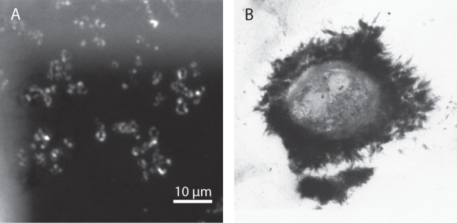
(A) Fluorescence image of Paracoccus denitrificans Pd 1222 grown in the presence of Fe(II). EPS was stained with WGA-Alexa Fluor 633 conjugate directly on TEM grids. Bright color indicates the fluorescing EPS shells. (B) TEM image of iron oxide precipitation around an encapsulated bacterium in a thin section collected at 432 m underground at the Äspö Hard Rock Laboratory near Oskarshamn, Sweden. The sample was stained with uranyl acetate to enhance the electron contrast of the biological material.