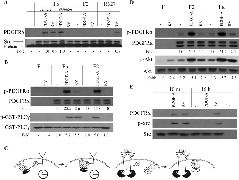
FIG 1.
RV induced a unique SFK-PDGFRα relationship, which was required for activation of PDGFRα and downstream signaling events. (A) Near-confluent, serum-starved Fα, F2, and R627 (a kinase inactive point mutant [74]) cells were stimulated with PDGF-A (50 ng/ml) or normal rabbit vitreous (RV) for 10 min. Where indicated, the cells were pretreated with vehicle or SU6656 for 30 min. The cells were lysed, the lysates were immunoprecipitated with an anti-Src antibody, and the resulting samples were subjected to Western blotting using an anti-PDGFRα antibody (27P) and an anti-Src antibody. The fold values are the PDGFRα/Src ratio or the p-Akt/Akt ratio. The position of the 50-kDa heavy chain of the immunoprecipitating antibody (H-chain) is indicated. The data presented are representative of three independent experiments. (B) Serum-deprived F, Fα, and F2 cells were treated with PDGF-A (50 ng/ml) or RV for 10 min and lysed, and PDGFRα was then immunoprecipitated with an anti-PDGFRα antibody (27P). The immunoprecipitates were subjected to an in vitro kinase assay in which the substrate was a GST-PLCγ fusion protein. The extent of phosphorylation was monitored by Western blotting using an antiphosphotyrosine antibody (pY20). The membranes were stripped and reprobed with antibodies against PDGFRα or GST. The experimental results presented are representative of three independent experiments. p-PDGFRα, phosphorylated PDGFRα. (C) (Left) SFKs act upstream of PDGFR; ROS-activated SFKs phosphorylate monomeric PDGFRα and subsequently associate with it. The kinase activity of SFKs is required for the formation of the complex. (Right) SFKs act downstream of PDGFR; PDGF dimerizes PDGFRs and thereby triggers autophosphorylation that enables stable association of SFKs. The kinase activity of PDGFRα is required for formation of the complex. P, phosphate group. (D) Serum-starved F, F2, and Fα cells were stimulated with PDGF-A (50 ng/ml) or RV for 10 min. Their lysates were subjected to Western blot analysis using the following antibodies: anti-PDGFRα pY742 for p-PDGFRα and anti-Akt pS473 for p-Akt. The fold values are the p-PDGFRα/PDGFRα ratio or the p-Akt/Akt ratio. The data presented are representative of three independent experiments. F cells express no PDGFRs. (E) Serum-deprived Fα cells were treated with PDGF-A (50 ng/ml) or RV for 10 min (10 m) or 16 h and lysed, and the resulting lysates were immunoprecipitated with an anti-Src antibody (mouse origin) or a nonimmune IgG as a control (C). The resulting immunoprecipitates were subjected to Western blot analysis using antibodies against phospho-Src or PDGFRα. The membrane was reprobed with an anti-Src antibody. The data presented are representative of three experiments.

