FIG 9.
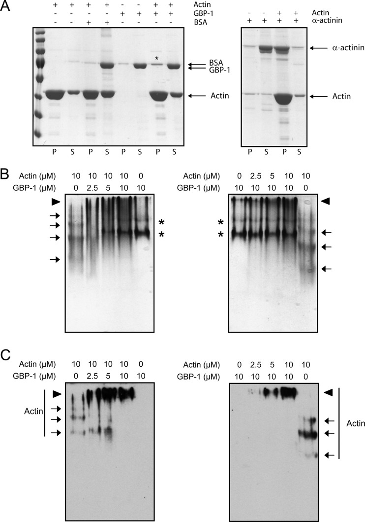
GBP-1 binds to both G-actin and F-actin. (A) F-actin was cosedimented by ultracentrifugation with GBP-1, BSA as a negative control, or α-actinin as a positive control. Equal amounts of pellets (lanes P) and supernatants (lanes S) were separated by SDS-PAGE. Gels were stained with Coomassie blue. An increase of GBP-1 in the pellet fraction was observed in the presence of F-actin, as indicated by an asterisk. (B) G-actin was incubated with GBP-1, and proteins were separated by native polyacrylamide gel electrophoresis. Gels were stained with Coomassie blue. Arrows, actin, which formed a ladder when incubated alone, indicating partial polymerization; asterisks, GBP-1 in its monomeric or dimeric form; arrowheads, actin–GBP-1 complexes. (C) Actin was detected using a specific antibody in a subsequent Western blot. Arrows, corresponding actin bands; arrowheads, larger protein complexes.

