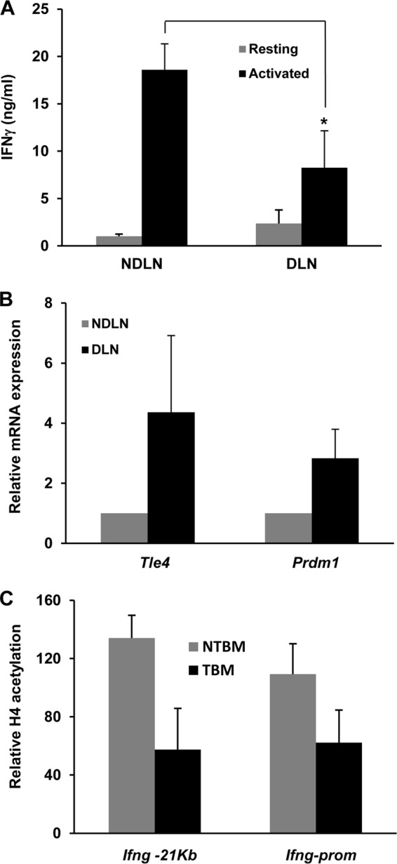
FIG 7.

Tumor-specific CD4+ T cells exhibit increased Tle4 and Blimp1 expression as well as reduced IFN-γ production. (A) IFN-γ expression in T cells isolated from draining (DLN) and nondraining (NDLN) lymph nodes from B16-OVA melanoma-bearing OT-II mice and stimulated for 18 h with T cell-depleted splenocytes loaded with OVA323–339 peptide, as measured by ELISA. Data are means and SEM for 5 different experiments. *, P < 0.05. (B) Similarly isolated CD4+ T cells from tumor-bearing mice were used to prepare total mRNA. Relative expression levels of mRNAs for Tle4 and Prdm1 were determined by qPCR. Bars represent relative expression in T cells isolated from DLN compared to cells from NDLN, expressed as means and SEM for 6 different experiments. (C) ChIP experiments were carried out to assess H4 acetylation at the Ifng −21kb CNS and promoter in CD4+ T cells isolated from DLN of tumor-bearing mice and from tumor-free mice. Bars show relative H4 acetylation in T cells isolated from DLN compared to cells from NDLN in tumor-bearing (TBM) and non-tumor-bearing (NTBM) mice, expressed as means and SEM for 3 different experiments.
