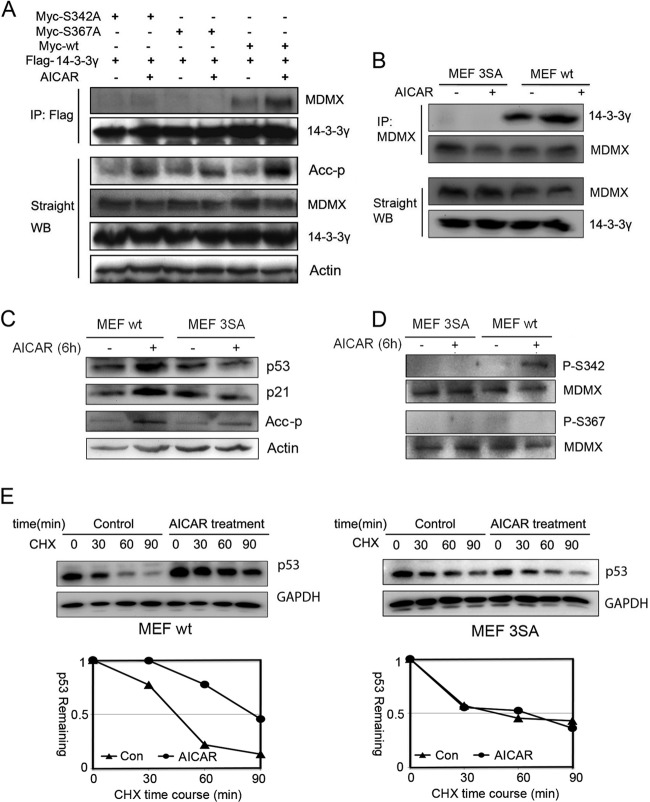
FIG 3.
Mutations of MDMX impair AMPK-mediated MDMX phosphorylation and p53 activation. (A) H1299 cells were transfected with the indicated plasmids. Five-hundred-microgram quantities of cell lysates were used for immunoprecipitation (IP) with anti-Flag antibodies, and precipitates were analyzed by WB with anti-Myc antibodies. Fifty-microgram quantities of cell lysates were loaded for straight WB analysis with the indicated antibodies. (B) Wild-type MEFs and MDMX-3SA mutant MEFs were treated with AICAR (1 mM) for 6 h. One-milligram quantities of cell lysates were used for IP with anti-MDMX polyclonal antibodies (Bethyl Laboratories), and precipitates were analyzed by WB with polyclonal anti-14-3-3γ antibodies (c-16). Eighty-microgram quantities of whole-cell lysates were analyzed by straight WB. (C and D) MEFs were treated with AICAR (1 mM) for 6 h before harvest. Eighty-microgram quantities of cell lysates were analyzed by WB with the indicated antibodies. (E) MEFs were treated with AICAR (1 mM) for 6 h, followed by CHX (50 μg/μl) treatment. Eighty-microgram quantities of cell lysates were analyzed by WB with the indicated antibodies. Quantitative results were analyzed with ImageJ software and plotted with Excel.

