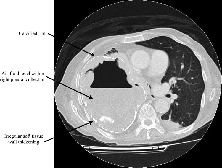
FIG 1.
Axial contrast-enhanced computerized tomography scan of the thorax at level of the pulmonary trunk displayed at the lung window showing a large right pleural collection with peripheral irregular soft-tissue densities, intermittent calcified rim, and air-fluid level within the collection (arrows).

