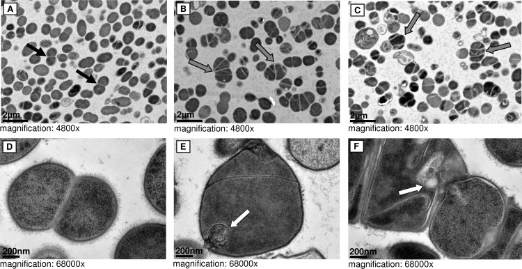
FIG 3.

Cell morphology. TEM pictures of the S. tigurinus parental strain 1366 (A and D) and SCV strains 2425 (B and E) and 2426 (C and F). Black arrows, regular cell separation; gray arrows, atypical and enlarged SCV cells with multiple cross walls; white arrows, irregularly shaped cells with mesosome-like structures.
