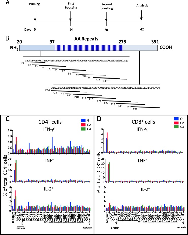
FIG 10.
Cell-mediated immunity of mice immunized with recombinant P. vivax CSP antigens. (A) C57BL/6 mice were immunized with three doses given 14 days apart, and IC expression assays were performed according to the timeline shown. (B) Twenty-six overlapping peptides spanning the entire sequence of the N or C terminus of PvCSP were used to stimulate splenocytes. AA, amino acid. (C and D) G1 mice (control) were primed i.m. at 2 × 10⁸ PFU/mouse with AdHu5-b-gal and boosted s.c. twice with poly(I·C). G2 mice were primed i.m. at 2 × 10⁸ PFU/mouse with AdC68-PvCSP and boosted twice with a protein mixture (His6-PvCSP-VK210, His6-PvCSP-VK247, and His6-PvCSP-Vivax-like, 30 μg/dose/mouse) in the presence of poly(I·C). G3 mice were primed and boosted with a protein mixture (His6-PvCSP-VK210, His6-PvCSP-VK247, and His6-PvCSP-Vivax-like, 30 μg/dose/mouse). Fourteen days later, splenic cells of these mice were cultured in the presence of anti-CD28 antibody, monensin, and brefeldin A with or without the peptides or recombinant proteins or ConA as indicated. The PvCS-VK210, PvCS-VK247, and PvCS-Vivax-like peptides represent the repeat regions of the allelic variants. After 12 h, cells were stained with anti-CD4, anti-CD8, anti-IL-2, anti-IFN-γ, and anti-TNF-α antibodies. The results are expressed as the total frequency (%) of CD4+ or CD8+ cells stained for each molecule. Results were obtained from pooled cells from five mice.

