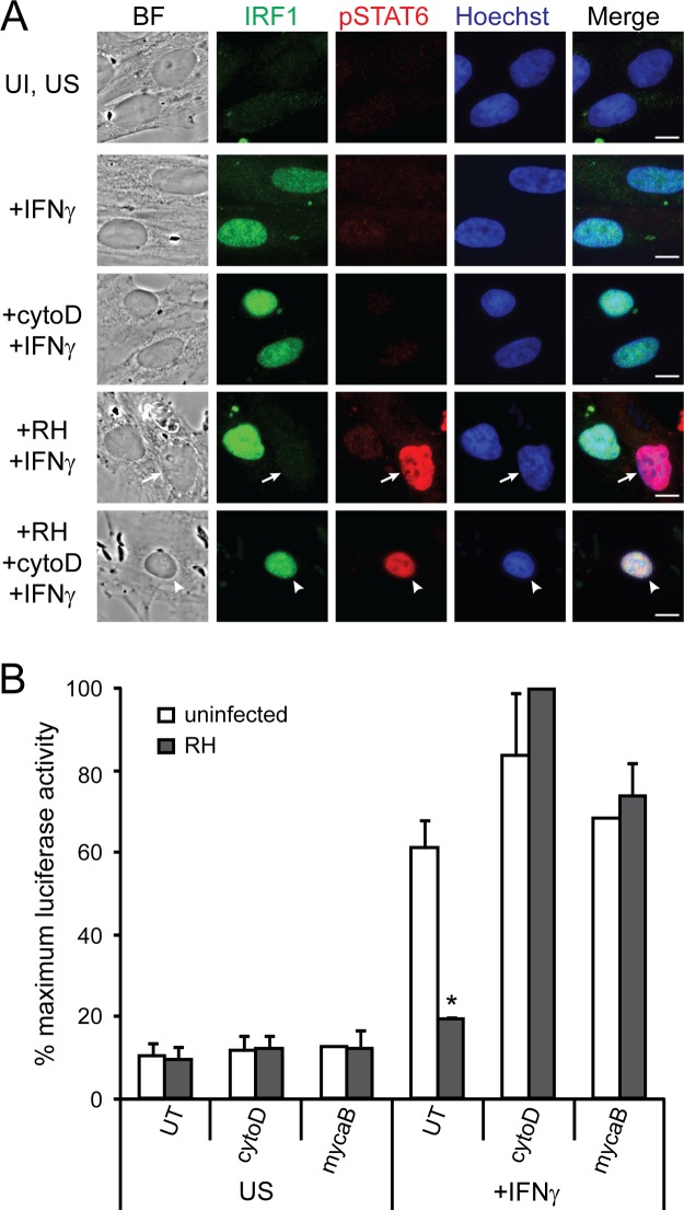
FIG 7.
Invasion is required for Toxoplasma's ability to inhibit IFN-γ-induced gene expression. RH parasites were pretreated with 1 μM cytochalasin D (cytoD) or 3 μM mycalolide B (mycaB) or left untreated (UT) and added to host cells for 1.5 h. Cells were then stimulated with 100 U of IFN-γ/ml or left unstimulated (US) for 18 h. (A) HFFs were fixed and stained for IRF1 (green), phospho-STAT6 (red), and with Hoechst dye (nucleus, blue). Scale bar, 10 μm. Arrows indicate infected cells, and arrowheads indicate uninfected cell with parasites attached and rhoptry proteins secreted. This experiment was performed three times with similar results. (B) A HEK293 GAS luciferase reporter cell line was then lysed, and the luciferase activity was measured. The results from two experiments per condition, except for the uninfected mycalolide B-treated condition for which only one experiment was done, were normalized to the maximum luciferase activity within the experiment and then averaged. In these experiments, 100% maximum induction represents an average of 10-fold induction over uninfected, unstimulated samples. Error bars represent the SEM. Asterisks (*) indicate P < 0.05 compared to uninfected control.

