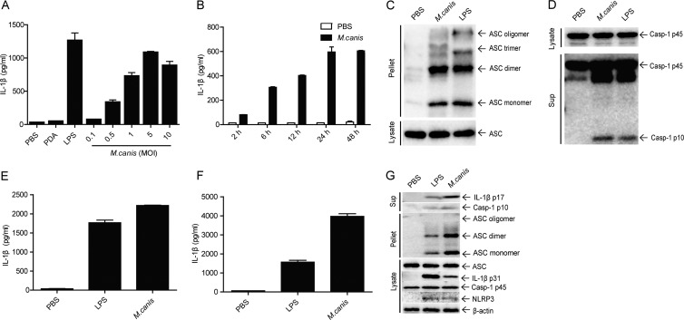
FIG 1.
M. canis-triggered IL-1β production and inflammasome activation in human monocytes, macrophages, and mouse dendritic cells. (A) THP-1 cells (1 × 10⁵) were infected with M. canis at different doses (MOIs), and 24 h later the supernatants were harvested for IL-1β measurement by ELISA. PBS- or PDA-treated cells served as negative controls. (B) THP-1 cells (1 × 10⁵) were treated with M. canis (MOI, 1), and supernatants were collected at different time points to detect IL-1β by ELISA. (C and D) THP-1 cells (1 × 10⁶) were incubated with PBS, M. canis (MOI, 1), or LPS (100 ng/ml) for 6 h, the cell pellets were collected for ASC pyroptosome detection, and the culture supernatants were harvested for caspase-1 assay via Western blotting. (E) THP-1-derived macrophages (1 × 10⁵) were treated with PBS, LPS (100 ng/ml), or M. canis (MOI, 1) for 6 h, and culture supernatants were collected for IL-1β detection via ELISA. (F) Mouse BMDCs (1 × 10⁵) were stimulated with PBS, LPS (1,000 ng/ml), or M. canis (MOI, 1) for 6 h, and the culture supernatant was collected for IL-1β detection via ELISA. (G) Mouse BMDCs (1 × 10⁶) were infected with M. canis (MOI, 1) or treated with PBS or LPS (1,000 ng/ml) for 6 h, and the culture supernatants were harvested for IL-1β immunoblotting. The cell pellets were used to detect ASC pyroptosome, and cell lysates were analyzed for detection of the expression of inflammasome components. Data in panels A, B, E, and F are means ± standard deviations from one out of three independent experiments. Data in panels C, D, and G are from one out of two independent experiments.

