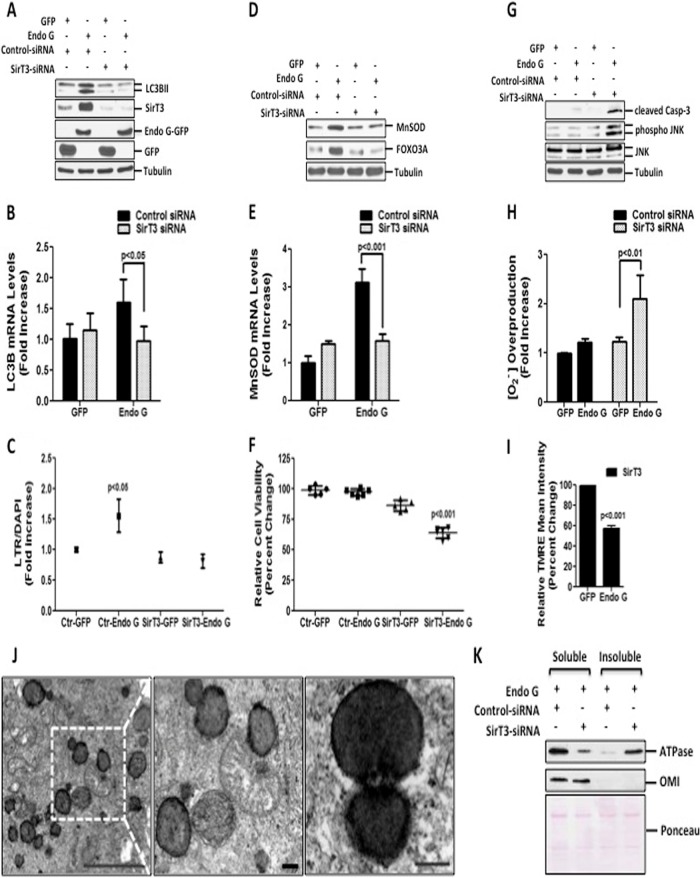
FIG 5.
SirT3 is required to maintain mitochondrial integrity and cellular viability of cells undergoing proteotoxic stress. (A) Lipidated levels of LC3B were detected by immunoblotting in cells transfected with 20 nM control siRNA against luciferase or against SirT3, followed by transfection with the indicated plasmids for 48 h. Endogenous levels of SirT3 and exogenous levels of Endo G-GFP and GFP were evaluated by Western blotting. (B) Endogenous mRNA levels of LC3B were determined by qRT-PCR of cells treated as described above for panel A. (C) LTR uptake was assessed in fixed cells treated as described above for panel A. The same results were obtained by using two different siRNAs against SirT3. (D) Immunoblots of crude lysates from cells treated as described above for panel A were used to detect the levels of the indicated proteins by probing with the respective antibodies. (E) Endogenous mRNA levels of MnSOD were determined by qRT-PCR of cells treated as described above for panel A. (F) Viability of MDA-MB231 cells, transfected as described above for panel A, was determined at 48 h. (G) Crude cellular lysates, treated as described above for panel A, were used to assess cleaved caspase-3, phosphorylated JNK, and total JNK levels by Western blotting. (H) Mitochondrial superoxide anion levels were determined by FACS analysis in cells transfected as described above for panel A and stained with MitoSox Red. (I) Mitochondrial potential was evaluated by FACS analysis in cells treated with 20 nM siRNA against SirT3 followed by transfection with either GFP or Endo G and stained with TMRE dye at 48 h. (J) Representative electron micrographs of mitochondria from MDA-MB231 cells transfected with 20 nM siRNA against SirT3 followed by Endo G-GFP at 48 h (scale bar, 5 μm). Also shown is a higher-magnification view of the selected region indicating fragmented mitochondria with electron-dense aggregates (scale bar, 0.5 μm). (K) The soluble and insoluble fractions of isolated mitochondria from cells transfected as indicated for 24 h and lysed with buffer containing Triton X-100 were subjected to Western blotting to detect FoF1 ATP synthase α subunit levels. Ponceau S staining served as a loading control.

