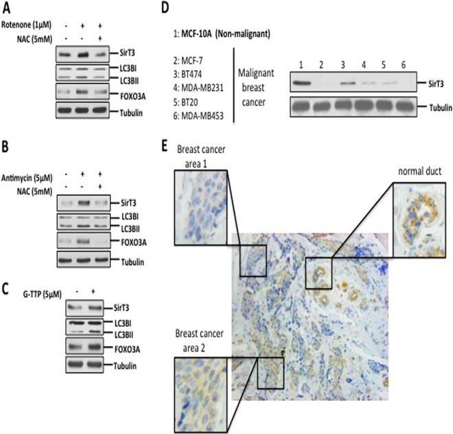
FIG 6.
SirT3 induction is triggered by other mitochondrial stressors and detected in human breast adenocarcinoma. (A and B) Crude extracts from cells treated for 24 h with 1 μM rotenone or 5 μM antimycin A in the presence or absence of 5 mM NAC were subjected to Western blotting and evaluated for levels of SirT3, FOXO3A, and lipidation of LC3B. (C) Lysates of cells treated with 5 μM G-TPP for 4 h were tested to determine the levels of the indicated proteins. (D) Crude lysates from the indicated cells were evaluated to determine SirT3 levels. (E) Representative histological image of a primary breast adenocarcinoma tumor tissue stained for SirT3. Higher-magnification views of the boxed region are shown.

