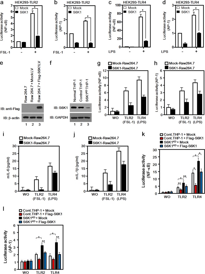
FIG 1.
S6K1 is negatively involved in the activation of NF-κB and AP-1 induced by TLR2 and TLR4. (a to d) HEK293-TLR2 cells (a and b) and HEK293-TLR4 cells (c and d) were cotransfected with a mock or Flag-S6K1 vector together with pBIIx-luc (a and c) or with AP-1-luc together with the Renilla luciferase vector (b and d). Twenty-four hours after transfection, cells were treated with or without FSL-1 (a and b) or LPS (c and d) for 6 h and then analyzed for luciferase activity. Results are expressed as the fold induction in luciferase activity relative to that in untreated cells. (e) The mock or Flag-S6K1 lentivirus vector was used to infect Raw 264.7 cells as described in Materials and Methods. The expression of Flag-S6K1 was examined with anti-Flag antibody by using Western blot assay. Immunoblotting (IB) with anti-β-actin antibody was performed to generate a control for gel loading. (f) A total of 2 × 10⁵ THP-1 cells were cultured in a 24-well plate and infected with lentiviral particles containing shRNA targeting human S6K1 (S6K1KD THP-1) or control shRNA lentiviral particles (Control THP-1). Cells were cultured in medium containing puromycin (4 to 8 μg/ml) for 2 weeks to select stable clones. The knockdown efficacy was examined with anti-S6K1 antibody using Western blotting. Immunoblotting with anti-GAPDH antibody was performed to generate a control for gel loading. (g and h) Mock-Raw 264.7 and S6K1-Raw 264.7 cells were cotransfected with pBIIx-luc (g) or AP1-luc (h) reporter together with Renilla luciferase vector. Twenty-four hours after transfection, cells were treated with or without (WO) FSL-1 or LPS for 6 h and then analyzed for luciferase activity. Results are expressed as the fold induction in luciferase activity relative to that in untreated cells. The data shown are the averages of a minimum of three independent experiments, with error bars denoting standard deviations (±SD). (i and j) Mock-Raw 264.7 and S6K1-Raw 264.7 cells were treated with or without TLR2 or TLR4 agonist (FSL-1 and LPS, respectively) for 9 h, and then supernatants were harvested. Levels of mouse IL-6 and mouse IL-1β were measured in the supernatants according to the assay manufacturer's protocol. (k and l) Control THP-1 and S6K1KD THP-1 cells were transfected with the mock or Flag-S6K1 vector with pBIIx-luc (k) or AP1-luc (l) together with Renilla luciferase vector. Twenty-four hours after transfection, cells were treated with or without FSL-1 or LPS for 6 h and then analyzed for luciferase activity. Results are expressed as the fold induction in luciferase activity relative to that in untreated cells. The data shown are the averages of a minimum of three independent experiments, with error bars denoting ±SD. *P < 0.01, **P < 0.05.

