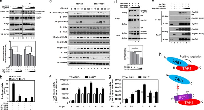
FIG 4.
S6K1 competitively interferes with the binding of TAB1 to TAK1, which inhibits TAK1 kinase activity. (a) HEK293 cells were transfected with Myc-TAK1 and different concentrations of Flag-S6K1. At 36 h after transfection, cells were treated with or without LPS (100 ng/ml) for 45 min, extracted, and Western blotting was performed with the antibodies indicated to the left. The band intensity of pho-TAK1 was analyzed with Image J (bottom). Data shown are the averages from a minimum of three independent experiments (±SD). *, P < 0.05. (b) HEK293-TLR4 cells were cotransfected with wt S6K1, DN-S6K1, or CA-S6K1 vector, together with pBIIx-luc reporter and Renilla luciferase vector. Twenty-four hours after transfection, cells were treated with or without (WO) LPS for 6 h and then analyzed for luciferase activity. Results are expressed as the fold induction in luciferase activity relative to that in untreated cells. The data shown are the averages of a minimum of three independent experiments, with error bars denoting ±SD. (c) Wild-type and S6K1KD THP-1 cells were treated with or without LPS for different times, and then Western blotting was performed as described in Materials and Methods. (d) HEK293 cells were transfected with Myc-TAK1, Flag-S6K1, and different concentrations of Flag-TAB1 as indicated. At 36 h after transfection, cells were extracted and immunoprecipitated with anti-Myc antibody. Interaction was detected by Western blotting with anti-Flag antibody. The presence of Myc-TAK1, Flag-S6K1, and Flag-TAB1 in the pre-IP lysates was verified by Western blotting. The band intensity of Flag-S6K1 was analyzed with Image J (bottom). The data shown are the averages of a minimum of three independent experiments (±SD). *, P < 0.05. (e) HEK293 cells were transfected with Myc-TAK1, Flag-TAB1, and different concentrations of Flag-S6K1 (66-333) as indicated. At 36 h after transfection, cells were extracted and immunoprecipitated with anti-Myc antibody. The interaction was detected by Western blotting with anti-Flag antibody. The presence of Myc-TAK1, Flag-TAB1, and Flag-S6K1 (66-333) in the pre-IP lysates was verified by Western blotting. (f and g) Wild type and S6K1KD THP-1 cells were treated with or without LPS (f) or FSL-1 (g) for different times as indicated. The kinase assay for TAK1 was performed using a c-TAK1 kinase assay kit in accordance with the manufacturer's protocol. The data shown are the averages of a minimum of three independent experiments (±SD). *, P < 0.05; **, P < 0.01. (h) A model for the negative regulation of TAK1. The C terminus of TAB1 interacts with the N terminus of TAK1, and the TAB1 association to TAK1 positively regulates TAK1 activity via the recruitment of p38 and induction of catalytic activity (top). In contrast, the interaction between the internal catalytic domain of S6K1 and the N terminus of TAK1 inhibits the TAB1 interaction with TAK1, which results in the inhibition of TAK1 catalytic activity.

