
FIG 1.
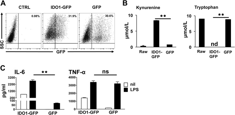
IDO enhances IL-6 production in macrophages. (A) RAW 264.7 macrophages were transduced with a lentivirus encoding an IDO1-GFP fusion protein, and 72 h later, IDO expression was assessed by flow cytometry as a function of GFP+ cells. Panels are representative of at least 5 samples per group. (B) Spontaneous kynurenine production and Trp consumption in IDO+ and control macrophage culture supernatants were assessed by HPLC as described in the text. (C) IDO+ and GFP+ control cultures were stimulated with LPS (1 μg/ml) for 18 h, and culture supernatants were tested by ELISA to determine concentrations of IL-6 and TNF-α. Bars represent the mean values for triplicate samples ± the standard deviations. **, P < 0.01 as determined by Student's t test. ns, not significant. Experiments were repeated at least three times, with similar results.