
FIG 4.
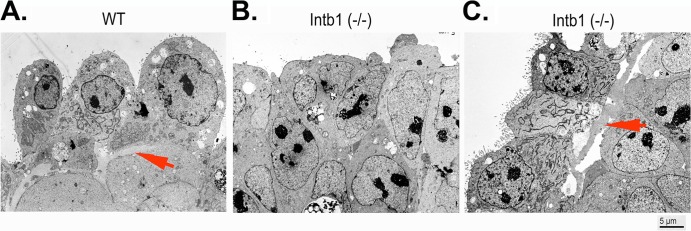
Analysis of embryoid bodies/spheroids using electron microscopy. Embryoid bodies (5 days in suspension culture) were analyzed by transmission electron microscopy. (A and B) Representative images are shown for surface layers of cells for wild-type (WT) (arrow indicates basement membrane) (A) and for β1 integrin-null [Intb1 (−/−)] (B) cells. (C) In some surface areas of the β1 integrin-null embryoid bodies, microvillus-containing endoderm cells are present, and loosely attached basement membranes are observable (arrow indicates basement membrane).