FIG 5.
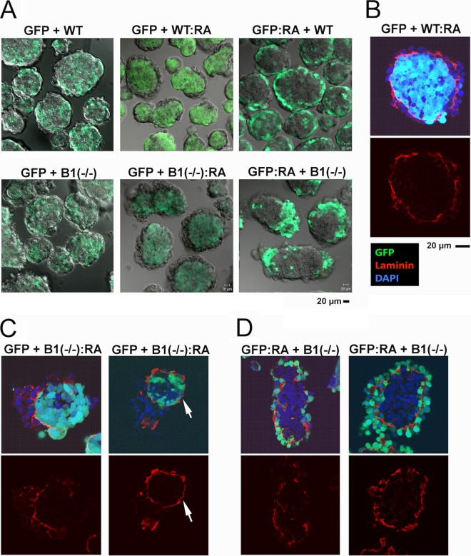
Analysis of chimeric embryoid bodies derived from β1 integrin-positive and -deficient ES cells. Wild-type ES cells expressing GFP (GFP), with or without differentiation by retinoic acid (1 μM for 7 days), were mixed with the β1 integrin-heterozygous G119 (designated “WT”) and the null G201 [B1(−/−)] ES cells with or without differentiation. The chimeric cell mixtures were allowed to aggregate and sort in suspension culture for 2 days. The time period was adequate for cell sorting but insufficient to induce spontaneous differentiation (29, 33). (A) The live chimeric embryoid bodies formed were analyzed for cell sorting patterns. Confocal GFP images of embryoid bodies in the middle section are shown. “GFP + B1(−/−):RA” refers to GFP-labeled wild-type ES cells mixing with β1 integrin-null ES cells that have been differentiated with retinoic acid. “WT” refers to unlabeled G119 β1 integrin-heterozygous ES cells. (B) A representative image of a chimeric embryoid body consisting of GFP-labeled wild type (GFP) mixed with retinoic acid-differentiated β1 integrin-heterozygous (WT:RA) ES cells that was stained for laminin (red), GFP (green), and DAPI (blue). Overlaying GFP and DAPI produces a pale blue color. (C) The embryoid bodies were produced from a mixture of the undifferentiated GFP-labeled and the differentiated β1 integrin-null ES cells [GFP + B1(−/−):RA]. Two examples show confocal images of GFP (green), laminin (red), and DAPI (blue). (D) Embryoid bodies were produced from a chimeric mixture of the differentiated GFP-labeled wild-type and the undifferentiated β1 integrin-null ES cells [GFP:RA + B1(−/−)]. Two examples of confocal images of GFP (green), laminin (red), and DAPI (blue) are shown.

