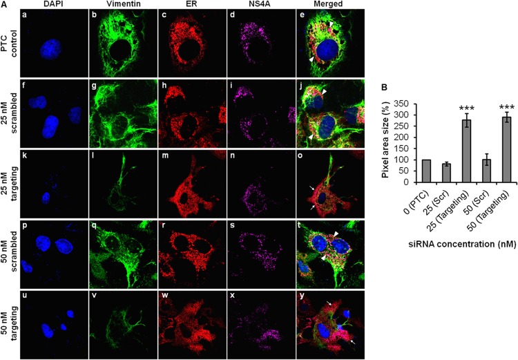
FIG 10.
Vimentin is essential for anchorage of DENV replication complexes mediated by NS4A interaction. (A) Huh-7 cells were treated with vimentin targeting or scrambled siRNA and fixed after DENV-2 infection for immunofluorescence analysis. The subcellular localization of vimentin (green), ER (red), and DENV NS4A protein (violet) were visualized by LSCM, with the nuclei stained with DAPI (blue). The images were taken at a magnification of ×100. White arrowheads denote the punctuate ER staining (concentrated localization), and white arrows denote diffused ER staining (dispersed localization). (B) Quantification analysis of the distribution of immunofluorescence signals from the IFM images, shown as percentages of pixel area size which made reference to the PTC. Statistical analyses were performed using one-way ANOVA and Dunnett's test (GraphPad Software). Values that are significantly different (P < 0.001) are indicated by three asterisks. Immunofluorescence signals were quantitated on a per-cell basis using the ImageJ software program. The number of cells counted per sample is 10. Values are means ± SD (error bars).

