
FIG 4.
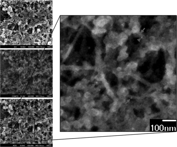
Ultrastructural studies illustrate the close association of vimentin and NS4A. Huh-7 cells were infected for 48 h and then subjected to detergent extraction, chemical fixation, and immunogold labeling for electron microscopic visualization of vimentin cytoskeleton. The enlarged image (panel iii) represents the overlay of the secondary electron image (panel i) and the backscattered electron image (panel ii). A close association of vimentin (gray arrows) with NS4A (gray arrowheads) is shown. Magnification, ×50,000. Bars = 100 nm.