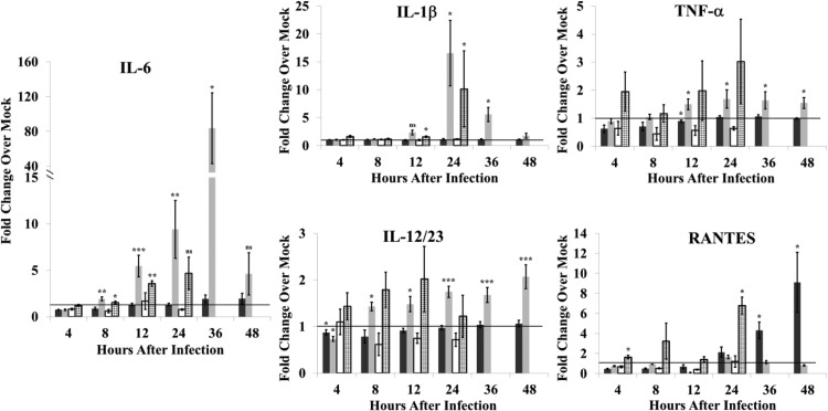
FIG 4.
Proinflammatory cytokine production in response to SHFV infection by MΦs isolated from disease-resistant and disease-susceptible NHPs. Fluids from MΦ cultures that were mock infected, infected with SHFV (MOI of 1.0), or incubated with UV-inactivated SHFV virus were analyzed for proinflammatory cytokines by multiplexed ELISA. Values shown represent average fold change in cytokine level in an SHFV-infected culture fluid compared to that in an autologous time-matched mock-infected culture fluid. The data were averaged from 20 baboon cultures infected with SHFV (black bars), 21 macaque cultures infected with SHFV (gray bars), 3 baboon cultures incubated with UV-inactivated SHFV (white bars), or 3 macaque cultures incubated with UV-inactivated SHFV (stippled bars). Error bars represent the standard error of mean. *, P ≤ 0.05; **, P ≤ 0.01; ***, P ≤ 0.005; ns, not significant.

