FIG 4.
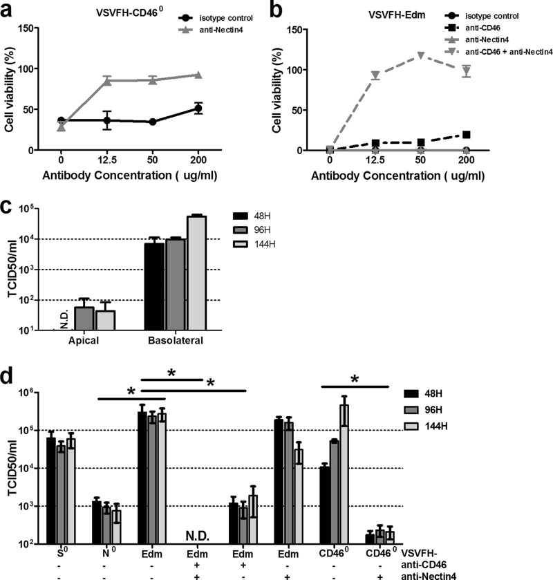
Effect of nectin4 ablation on viral shedding. Calu-3 cells in the 96-well plate were infected with VSVFH-CD460 (a) or VSVFH-Edm (b) in the presence of monoclonal antibodies specifically against human nectin4 and/or CD46 or an isotype control at the indicated concentrations. Cell viability was assessed at 48 h postinfection using the MTS cell proliferation assay. (c) Evaluation of virus shedding in vitro using the Calu-3 human airway epithelial model. Polarized layers of Calu-3 cells on 12-well Transwell filters were infected with VSVFH-Edm from either the apical or basolateral surface of airway epithelial cells as indicated on the x axis. Supernatants were collected from either basolateral chambers (apical infection) or apical chambers (basolateral infection) at the indicated time points. (d) Calu-3 cells on 12-well Transwell filters were infected with VSVFH-N0, VSVFH-S0, VSVFH-CD460, or VSVFH-Edm basolaterally in the absence or presence (200 μg/ml) of antibodies as indicated below the graph. The amounts of infectious viral particles released to the supernatant obtained from apical chamber at 48 h, 96 h, and 144 h postinfection were determined by TCID50 assays on Vero-SLAM cells. Results are expressed as the means ± SDs of three different experiments. *, P < 0.05 (unpaired student t test). N.D., not detectable.

