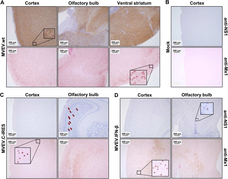
FIG 7.
Virus and Mx1 protein accumulation in the brains of MVEV-infected mice. Adult BALB.A2G-Mx1 mice were intracranially inoculated with 104 PFU of MVEV.wt (mouse B1-4) (A), diluent alone (mock, mouse A1-1) (B), MVEV.C-IRES (mouse C1-4) (C), and MVEV.IFN-β (mouse D1-4) (D). Mice were killed at 4 days p.i., 4-μm sagittal brain sections were prepared, and neighboring sections were immunostained for the virus protein NS1 (anti-NS1) or Mx1 (anti-Mx1). Sections were then counterstained using either hematoxylin (together with anti-NS1) or eosin (with anti-Mx1). Insets show the typical cytoplasmic and nuclear staining patterns of NS1 and Mx1, respectively. Arrows point to a number of NS1-positive cells in the olfactory bulb of mouse C1-4. Bars, 200 μm.

