
FIG 6.
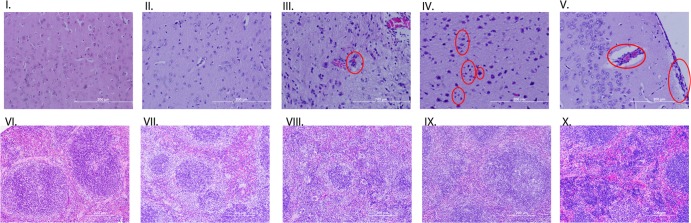
Histopathology staining of brain and spleen tissues. Sections I and VI are from an uninfected IFN-αβ/γ R−/− mouse showing no neuronal death and good spleen structure. Sections II and VII are from a C57BL/6 mouse infected with MACV with no visible neuron death or inflammation and good spleen structure. Sections III and VIII are from an IFN-αβ/γ R−/− mouse euthanized at 14 dpi, the red circle identifying vascular infiltrates. Sections IV and IX are from an IFN-αβ/γ R−/− euthanized at 24 dpi; red circles identify microglial cells and cellular debris from dead neuronal cells. Sections V and X are from an IFN-αβ/γ R−/− mouse that died at 34 dpi; red circles identify increased vascular and perivascular cellular infiltrates.