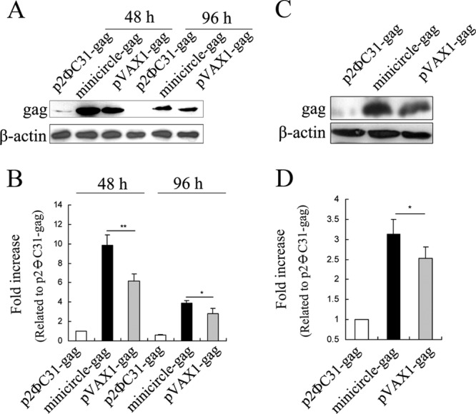
FIG 2.

Determination of HIV-1 gag gene expression mediated by minicircle DNA. (A) C2C12 cells were transfected with 2.0 μg of p2ΦC31-gag, 2.0 μg of pVAX1-gag, or 1.14 μg of minicircle-gag (equimolar with pVAX1-gag) and harvested at 48 h and 96 h posttransfection, respectively. Expression of gag was monitored by Western blotting. (C) BALB/c mice were intramuscularly injected with 20.0 μg of p2ΦC31-gag, 20.0 μg of pVAX1-gag, or 11.4 μg of minicircle-gag (equimolar with pVAX1-gag). The samples were harvested 7 days later for Western blot analysis. (B and D) The histograms indicate the levels of the protein determined from 3 independent experiments expressed as the fold change relative to that in the p2ΦC31-gag control after normalization to β-actin. Values are means ± SDs. *, P < 0.05 versus the pVAX1-gag control.
