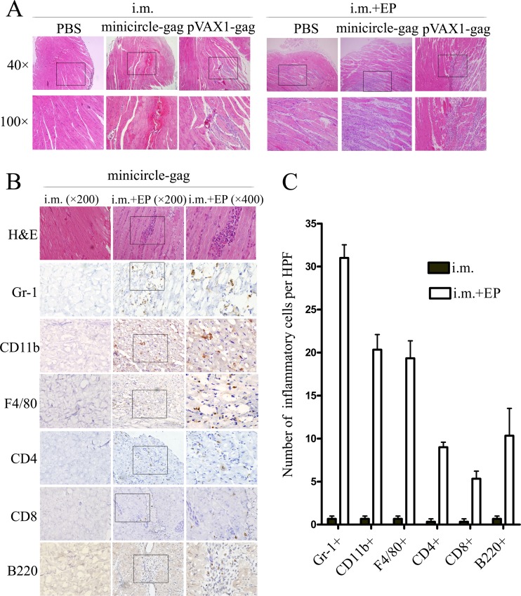
FIG 5.
Immune cell infiltration at the i.m. injection site induced by EP. Groups of five BALB/c mice were injected with PBS, 20 μg of minicircle-gag, or 20 μg of pVAX1-gag i.m. with or without EP. Four days later, the injected muscles were obtained for H&E staining (A) and immunohistochemical analysis (B) with the antibodies as indicated. (C) The mean numbers of inflammatory cells per high-power field (HPF) (×200). Data shown are representative of three independent experiments.

