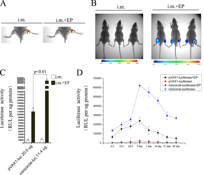
FIG 7.
Detection of the biodistribution and expression of minicircle DNA delivered i.m. with or without in vivo EP at the injection site. (A) BALB/c mice were injected with 20 μg of EMA-labeled minicircle-luciferase with or without EP. Ten minutes later, the biodistribution of minicircle DNA was determined using an in vivo imager. (B) BALB/c mice were injected with 20 μg of minicircle-luciferase with or without EP. Seven days later, the expression of luciferase was measured in vivo using an in vivo image system. (C) BALB/c mice were injected with 20 μg of pVAX1-luciferase, or 11.4 μg of minicircle-luciferase (equimolar with pVAX1-luciferase) i.m. with or without EP. At 7 days postinjection, the involved muscles were surgically removed; total cell lysates were prepared and analyzed with the luciferase assays. (D) BALB/c mice were injected as for panel C. The involved muscles were surgically removed at the indicated time points; total cell lysates were prepared and analyzed with the luciferase assays.

