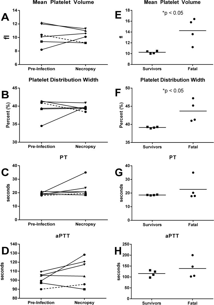
FIG 3.
Platelet and coagulation changes after infection with RVFV. Blood samples collected from AGM (A to D) and marmosets (E to H) were analyzed for changes in platelets and coagulation parameters. Graphs for AGM show differences between preinfection and at necropsy for the AGM that survived infection (dashed line) compared to AGM that succumbed (solid lines). Graphs for marmosets show differences between marmosets that survived or succumbed to infection at the time of necropsy.

