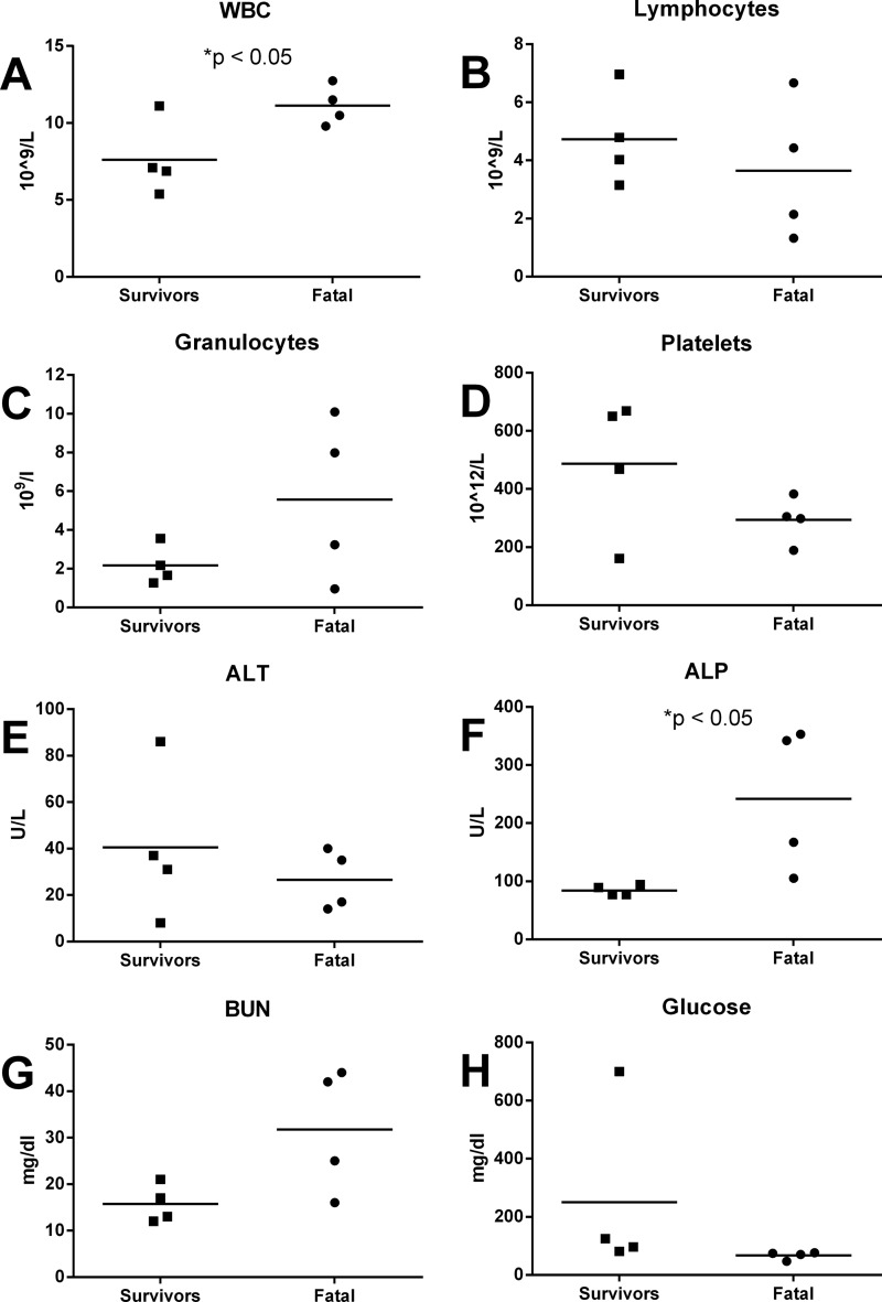
FIG 4.
Hematological changes in marmosets after infection with RVFV. Blood samples were collected at necropsy for marmosets exposed to aerosolized RVFV. Graphs show the results of CBC and serum chemistry analyses for individual marmosets that survived RVF exposure and marmosets that succumbed (symbols), with the averages (horizontal lines) for each group. (A) White blood cells (WBC); (B) lymphocytes; (C) granulocytes; (D) platelets; (E) alanine transaminase (ALT); (F) alkaline phosphatase (ALP); (G) blood urea nitrogen (BUN); (H) glucose.

