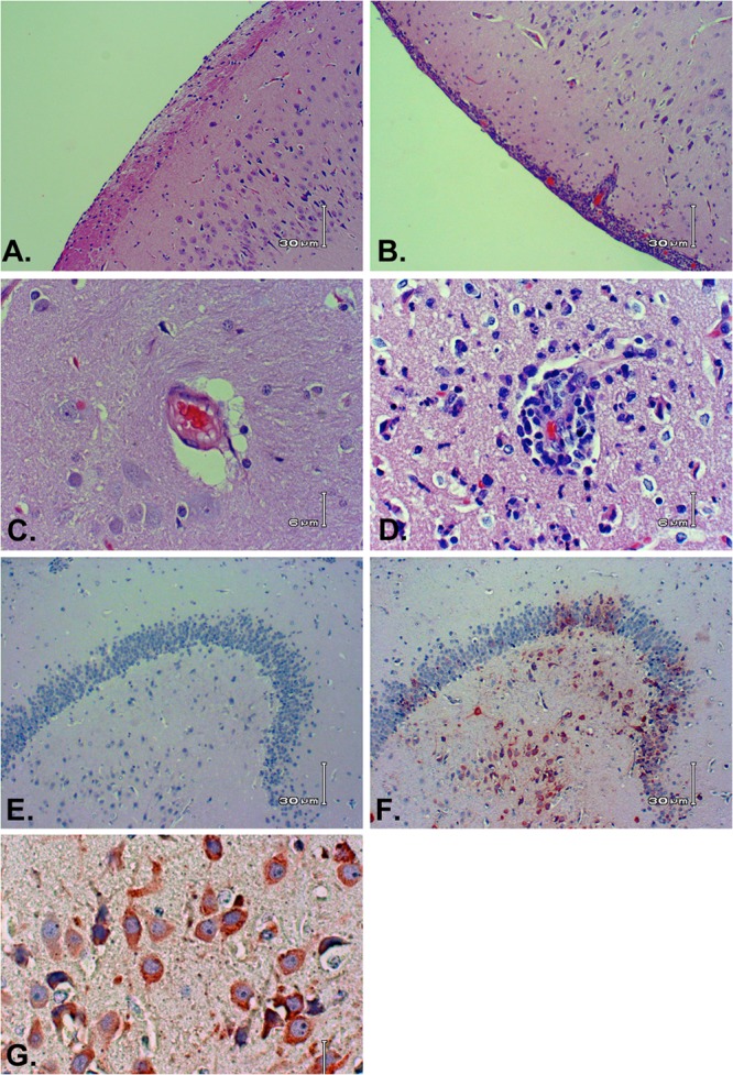
FIG 6.

Representative histologic images from marmosets infected with RVFV. Virus-infected brain showing normal meninges (A) compared to lymphocytic meningitis (B); virus-infected brain showing a normal blood vessel (C) compared to a vessel with perivascular lymphocytic inflammation (D); (E) virus-infected brain stained with nonimmune IgG (negative control); (F) same virus-infected brain showing RVFV immunostaining in neurons in the hippocampus; (G) RVFV-infected neurons at higher magnification.
