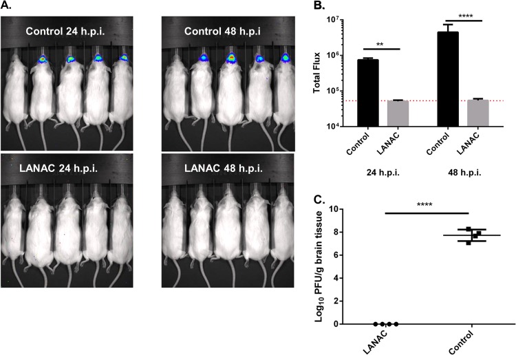
FIG 5.
In vivo bioluminescence imaging of the protective effects of LANACs. (A) Nonimmunized and E1ecto LANAC-immunized mice (n = 4/group) were challenged by i.n. inoculation with 104 PFU of WEEV.McM.FLUC virus and then imaged at 24 and 48 hpi. In each image, the first mouse on the left is an uninfected control to establish background bioluminescence. (B) Bioluminescence quantification. Each bar represents the average bioluminescence signal ± standard deviation for each treatment group. Differences between the two treatment groups were significant (P < 0.005). (C) Infectious virus titers in homogenates of brains isolated from animals used for panel A at 72 hpi. Differences between the two treatment groups were statistically significant (P < 0.005).

