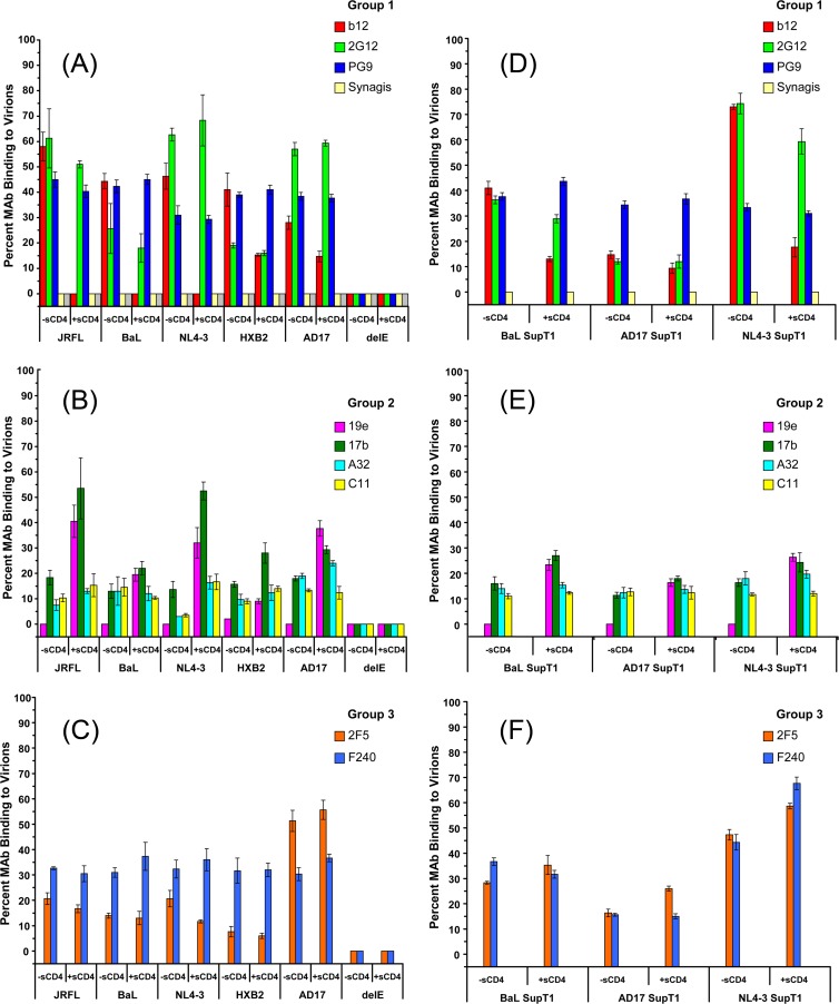
FIG 5.
Anti-envelope MAb binding to viral particles in solution. Viruses were produced in either HEK293T cells (A to C) or SupT1 cells (D to F) and tested at 10-μg/ml p24 equivalent concentrations with or without 100 μg/ml sCD4. The indicated groups of MAbs were tested at 4.5- to 6.6-μg/ml final concentrations (see Materials and Methods). Binding data for MAbs b12 and 17b from Fig. 4 with JRFL, BaL, and NL4-3 viruses are included in panels A and B, respectively. The relative fraction of MAb that adopts a lower diffusion coefficient (∼8 μm2/s) as a result of virion binding (see Materials and Methods) is shown. All experiments were repeated at least three times, and average values are shown. Error bars indicate standard deviations.

