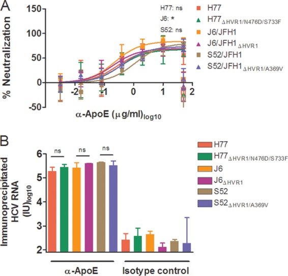
FIG 9.

ApoE-specific neutralization and ApoE HCV particle association were similar for viruses with and without HVR1. (A) H77, J6, or S52 HCV with and without HVR1 was incubated for 1 h at 37°C in four replicates with anti-ApoE MAb (1D7) in the indicated dilution series, along with four replicates with 50 μg/ml of control antibody (mouse IgG1) and 6 replicates with medium only. The previous day, Huh7.5 cells were plated in 96-well plates. These cells were incubated with the above-mentioned virus or virus-antibody mixtures, washed after 3 h, and incubated for an additional 45 h with complete medium prior to HCV staining. FFU counts at the given antibody dilutions were normalized to the mean FFU count of the 6 replicates of virus only. The data points are means and SD of four replicates. The open symbols represent the control antibody only tested at the highest antibody concentration. Four-parameter nonlinear curve regression was used to fit the data points. Z-tests were used to compare the differences in maximum attained blocking for each antibody. Pairwise comparisons were made between viruses of the same isolate. *, statistical significance at a P value of <0.05; ns, not statistically significant. (B) ApoE-specific immunoprecipitation was carried out as described in Materials and Methods using ApoE-specific antibody (1D7) or relevant IgG1 mouse control antibody, and the amounts of immunoprecipitated HCV RNA were measured by RT-qPCR in duplicate on the eluted fractions in a LightCycler with primers and protocols that have been described previously (29). The results are shown as the total amount of HCV RNA in each sample (cutoff, 80 IU). The error bars represent SD. t tests were performed comparing HCV RNA immunoprecipitation for HCV with and without HVR1. ns, not statistically significant.
