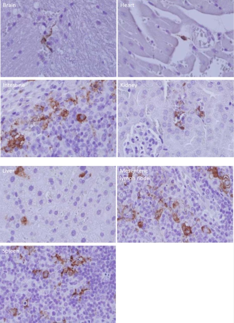
FIG 2.
Immunohistochemistry-stained tissues of a mouse infected with SFTSV. The anti-SFTSV antibody-stained cells are shown in each organ: an unidentified perivascular cell in the cerebrum; an unidentified perivascular, spindle-shaped cell in the myocardium; mononuclear cells of an intestinal submucosal lymphoid follicle; unidentified cells in the location of intertubular capillaries in the renal cortex; hepatocytes and sinusoidal lining cells; unidentified mononuclear cells in a mesenteric lymph node; and unidentified mononuclear cells in the spleen. The experiments were repeated 3 times, and the results were consistent.

