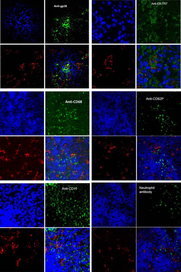
FIG 3.
Confocal microscopic images of thin sections of SFTSV-infected mouse spleen stained with antibody to gp38 and antibody to ER-TR7 recognizing reticular cells, antibody to CD68 recognizing macrophages, antibody to CD62P recognizing megakaryocytes/platelets, antibody to Ly-6 recognizing neutrophils, and antibody to CD11c (dendritic cells). Each panel shows the following: top left, DAPI-stained cell nuclei (blue); top right, specific antibody-stained cells (green); bottom left, rabbit anti-SFTSV antibody-labeled SFTSV (red); bottom right, merged images. SFTSV colocalized with gp38-stained host cells (yellow dots) and with antibody to ER-TR7-stained cells (SFTSV surrounding ER-TR7 markers inside cells). No colocalization of SFTSV with other cells was observed. The confocal microscopy for each antibody was repeated at least five times, and the results were consistent.

