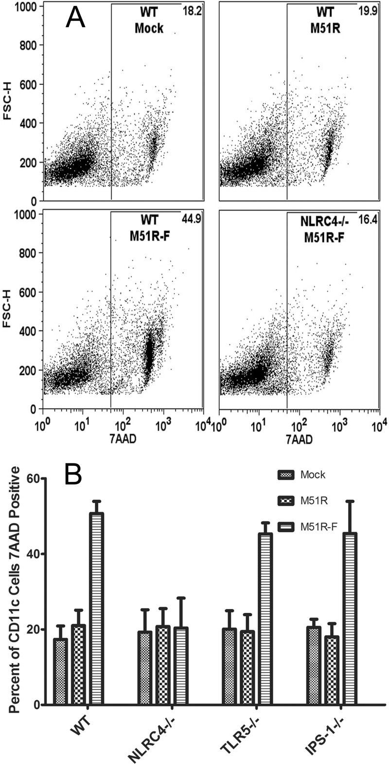
FIG 6.
Membrane permeabilization in DCs following viral vector treatment. DCs from WT, NLRC4−/−, IPS-1−/−, and TLR5−/− mice were treated with either M51R or M51R-F vector (MOI = 10) for 6 h. Cells were stained with 7AAD to measure membrane permeability and analyzed by flow cytometry. (A) Representative dot plots depicting forward scatter (FSC-H) versus 7AAD in arbitrary fluorescence units. Numbers indicate the percentage of positive cells in the gate. (B) Results of multiple experiments expressed as percentage of CD11c positive that were also 7AAD positive (means ± SD; n ≥ 3 experiments per group).

