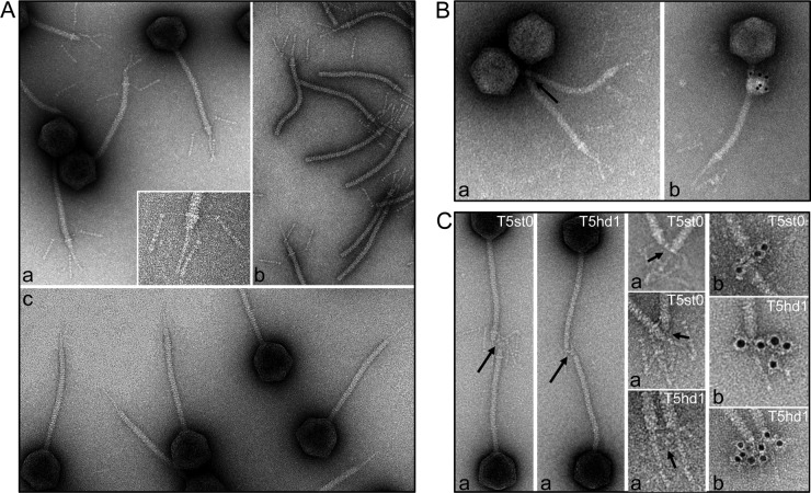
FIG 3.
(A) Analysis of the tail tip morphology in phage T5st0 in wide-field and blowup views (a), in isolated tails (b), and in the T5hd1 mutant lacking the L-shaped fibers (c). (B) Localization of T5p142 in T5st0 and (C) of pb3 in T5st0 and T5hd1: the position of the proteins was identified by IgG cross-linking (a, black arrows) or by visualization of the IgG molecules associated with goat anti-rabbit IgG–gold conjugate (b). For pb3, blowups of the tail tips are shown to highlight immunolocalization. The diameter of the tail tube is 12 nm.

