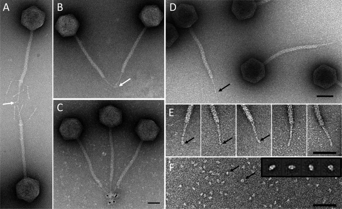
FIG 5.
Localization of pb5 in the tail tip. Immunolocalization of pb5 by IgG cross-linking (white arrows) on phages T5st0 (A) and T5hd1 (B) or by visualization of the IgG molecules associated with anti-rabbit IgG–5-nm gold conjugate on T5hd1 (C). Panel E shows a gallery of tail tips selected from high-resolution images of T5hd1 (D). Panel F shows an image of purified pb5 (black arrows) with a gallery of the major class averages obtained by processing 659 selected single particles of pb5 in the inset (23). Bars, 50 nm.

