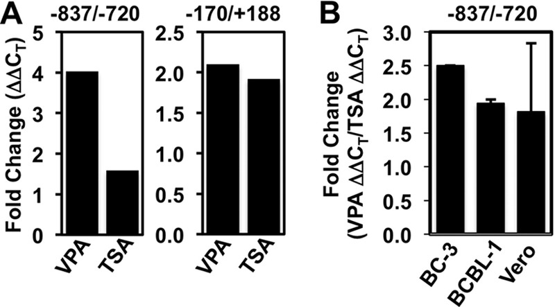
FIG 5.

VPA is slightly better than TSA in inducing histone acetylation on the Rta promoter. (A) BC-3 cells were treated with the indicated HDACi or mock treated. ChIP assays were performed using anti-H3K9/K14-ac and total rabbit IgG (negative-control) antibodies. Chromatin immunoprecipitated DNA was quantitated by real-time PCR using primer/molecular beacon sets (−837/−720) or SYBR green (−170/+188) specific for the indicated regions of the Rta promoter. Fold changes were calculated using the ΔΔCT method and represent the increased ChIP of H3-acetylated chromatin relative to that of IgG in the presence or absence of the inducing chemical. (B) The indicated KSHV-infected cells were treated with VPA or TSA or left untreated. ChIP assays were performed as described for panel A, and the fold changes in H3 acetylation at the −837/−720 Rta promoter element were determined. The fold changes for VPA and TSA were calculated individually using the ΔΔCT method and represent the increased ChIP of H3-acetylated chromatin relative to the ChIP of IgG in the presence or absence of the inducing chemical. Then, the fold increase induced by VPA was divided by the fold increase induced by TPA for each cell line and plotted. Data represent the fold increase of acetylation induced by VPA divided by that induced by TSA; the results in the BC-3 bar were calculated from the data shown in panel A.
