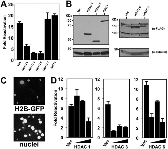
FIG 7.
Ectopic HDACs 1, 3, and 6 inhibit TPA-induced KSHV reactivation. (A) BCBL-1 cells were electroporated with 30 μg plasmids expressing the indicated HDACs or empty vector (Vec). Cells either were left untreated or were treated with TPA at 24 h postelectroporation. At 72 h posttreatment, reactivation was scored by measuring the percentage of cells expressing K8.1 by immunofluorescence; fold reactivation was scored by dividing the percentage of K8.1-positive cells transfected with each plasmid and treated with TPA by the percentage of identically transfected cells not treated with TPA. Cells were transfected with 30 μg of each plasmid. (B) Cellular protein extracts corresponding to the electroporations whose results are shown in panel A were tested by SDS-PAGE/Western blotting for expression of the indicated proteins. Asterisks in the right panel, ectopically expressed proteins. (C) BCBL-1 cells were electroporated with 30 μg histone H2b-GFP-expressing plasmid, fixed, and stained with DAPI (4′,6-diamidino-2-phenylindole) at 72 h postelectroporation. Using fluorescence microscopy, transfection efficiency was quantitated by dividing the number of GFP-positive cells by the total number of DAPI-positive cells. A representative field is shown in gray scale. (D) BCBL-1 cells electroporated with 10, 20, or 30 μg plasmids expressing the indicated HDACs were treated with TPA, and reactivation was scored as described for panel A.

