
FIG 5.
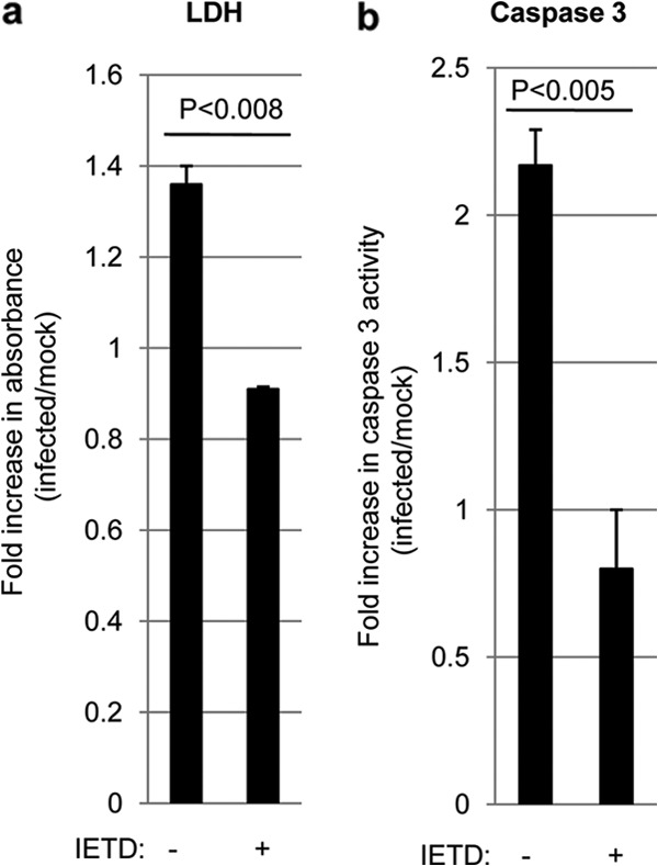
Inhibition of caspase 8 blocks WNV-induced apoptosis and tissue injury in an ex vivo model of WNV pathogenesis. Four 400-μm-thick tissue slices were prepared from individual brains extracted from 3-day-old SW mice. Slices were treated with 10 μM Z-IETD-FMK (IETD) for 24 h before being infected with WNV (105 PFU per slice). At 7 dpi, medium from the cultures was analyzed for the presence of lactate dehydrogenase (LDH). This medium contained any LDH released over the prior 2 days. The graphs show the fold increases in LDH levels (a) and caspase 3 activation (b) in WNV-infected BSC compared to mock-infected controls. Error bars represent the standard errors of the means.