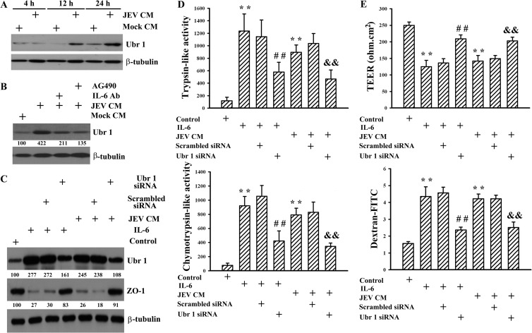
FIG 8.
Role of Ubr 1. Pericytes were mock infected (Mock CM) or infected with JEV (MOI, 20; JEV CM) for 48 h. The supernatants were collected and mixed with an equal volume of fresh medium. (A) The manipulated media were added to brain microvascular endothelial cells over time. Total cellular proteins were isolated and subjected to Western blotting with antibodies against Ubr 1 and β-tubulin. One representative blot of four independent experiments is shown. (B) Brain microvascular endothelial cells were exposed to the manipulated media (Mock CM and JEV CM) in the absence or presence of AG490 (50 μM) for 24 h. One set of manipulated medium (JEV CM) was modified by neutralization with IL-6 neutralizing antibody (10 μg/ml) for 30 min before being subjected to exposure. Total cellular proteins were isolated and subjected to Western blotting with antibodies against Ubr 1 and β-tubulin. One representative blot of four independent experiments is shown. The content in Mock CM was defined as 100%. Brain microvascular endothelial cells were transfected with control siRNA (1 nM) (mock transfection) or Ubr 1 siRNA (1 nM) for 4 h. The resultant cells were treated with IL-6 (20 ng/ml) or exposed to JEV CM for 24 h. Untreated cells were used as a control. (C) Total cellular proteins were isolated and subjected to Western blotting with antibodies against Ubr 1, ZO-1, and β-tubulin. One representative blot of four independent experiments is shown. The content in control was defined as 100%. (D) Cellular proteins were isolated and subjected to fluorogenic assay for determination of trypsin-like (upper graph) and chemotrypsin-like (lower graph) proteasome activities. n = 4. (E) The TEER (upper graph) and transendothelial permeability to dextran-FITC (lower graph) were measured. n = 4. **, P < 0.01 versus medium control; ##, P < 0.01 versus IL-6 control; &&, P < 0.01 versus JEV CM control.

