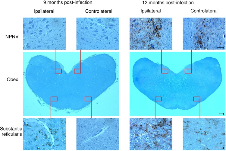
FIG 2.
Representative immunohistochemical patterns of PrPSc deposition within the brain at the obex level of preclinically scrapie affected sheep following intratonsillar inoculation. At 9 months p.i., the earliest PrPSc deposition was observed in the obex as glial-perivascular patterns in the substantia reticularis ipsilaterally to the inoculum side. The corresponding symmetrically contralateral area, as well as both the ipsilateral and contralateral nucleus parasympathicus nervi vagi (NPNV), were negative for PrPSc. At 12 months postinfection, a PrPSc deposition was found bilaterally in the obex at the level of both the NPNV and the substantia reticularis. Micrographs are higher magnifications of the red line-enclosed area of the transversal section of the obex (macrophotograph). Scale bars, 1,000 μm (macrophotograph) and 100 μm (micrographs).

