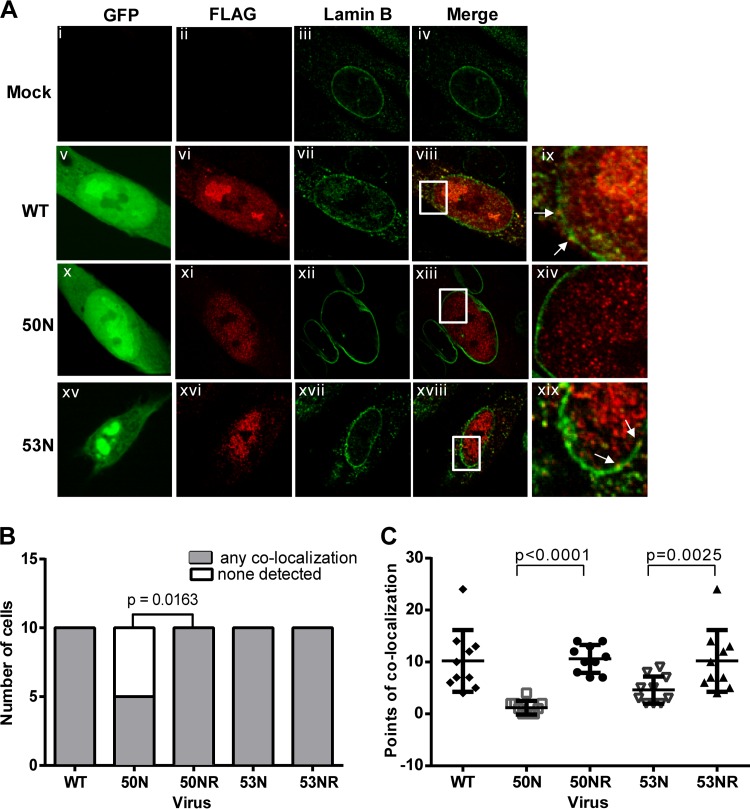
FIG 7.
Cellular distribution of UL97 in the absence of HCMV UL50 or UL53. (A) HFFs were mock electroporated (i to iv) or electroporated with WT (v to ix), UL50 null (50N) (x to xiv), or UL53 null (53N) (xv to xix) pBADGFP expressing FLAG epitope-tagged UL97. HFFs were also electroporated with rescued derivatives 50NR and 53NR. Cells were fixed on day 7 and stained with antibodies against FLAG (red) and lamin B (green). GFP-positive cells were located by confocal microscopy. Panels ix, xiv, and xix represent magnified (×3) sections of the insets in the panels to their left. White arrows point at regions of colocalization. (B) For statistical analysis, 10 infected (GFP-positive) cells from each electroporation were analyzed for whether they showed any points of colocalization (shaded bars) between FLAG-UL97 and lamin B or no observable colocalization (unshaded bars). The difference between the data for cells infected with 50N versus 50NR was analyzed using one-tailed Fisher's exact test, and the P value is shown. (C) The number of foci of FLAG-UL97 and lamin B colocalization was counted in representative optical sections of 10 cells/sample. Differences in these data between cells infected with a mutant versus its rescued derivative were analyzed using one-way analysis of variance (ANOVA) followed by Sidak's multiple-comparison test, and P values are shown.

