FIG 2.

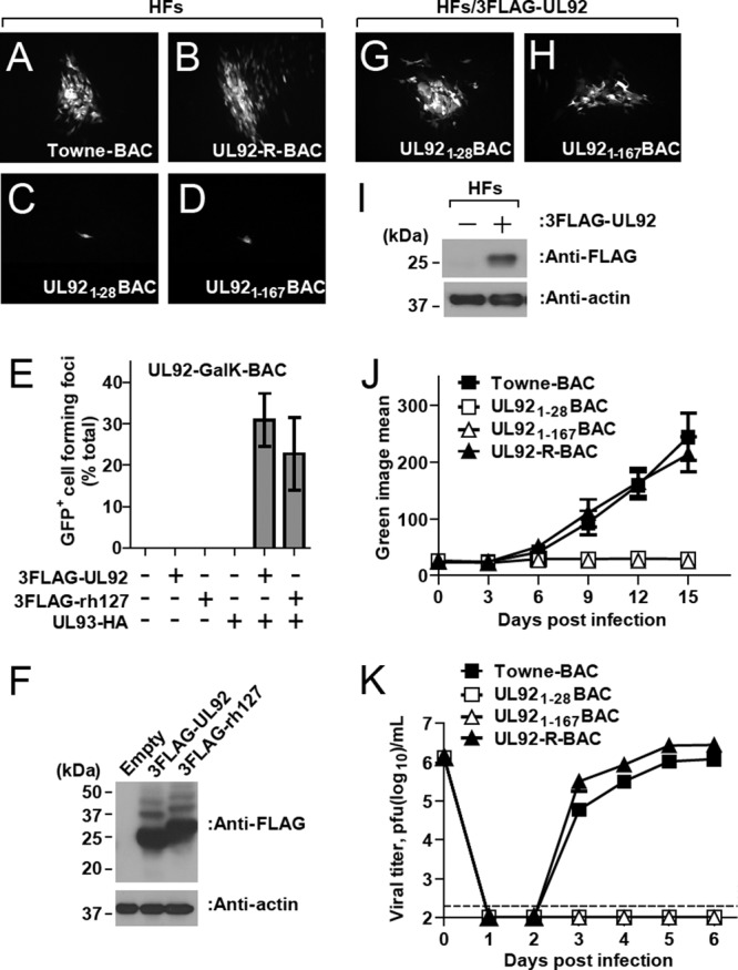
Replication properties of UL92 mutant BAC-derived viruses. (A to D) Typical GFP-positive plaque phenotypes at 10 days posttransfection of HFs with Towne-BAC (A), UL92-R-BAC (B), UL921-28BAC (C), and UL921-167BAC (D). Original magnification, ×100. (E) Secondary spread assay evaluation of plasmids 3FLAG-UL92 or 3FLAG-rh127 to complement UL92-GalK-BAC in the presence or absence of UL93-HA. (F) 293T cell lysates at 48 h posttransfection with indicated plasmids were subjected to immunoblotting with anti-FLAG antibody. The antibody against β-actin was used as a loading control. (G and H) Fluorescent images of GFP-positive cells at 10 days posttransfection of 3FLAG-UL92-expressing HFs with UL921-28BAC (G) and UL921-167BAC (H). Original magnification, ×100. (I) Lysates from 3FLAG-UL92-expressing HFs were subjected to immunoblotting with anti-FLAG antibody. The antibody against β-actin was used as a loading control. (J) Multistep growth curves (MOI of 0.01) of Towne-BAC and UL92 mutant viruses in HFs. Virus replication was quantified for 15 days based on the GFP signal intensity, which is expressed from a cassette within the viral genome. (K) Single-step growth curves (MOI of 3) of Towne-BAC- and UL92 mutant BAC-derived viruses in HFs. Virus titers were determined by plaque assays of supernatants collected at daily intervals. The data point at 0 dpi indicates the input virus dose. The detection limit of the plaque assay is indicated by a dashed line.
