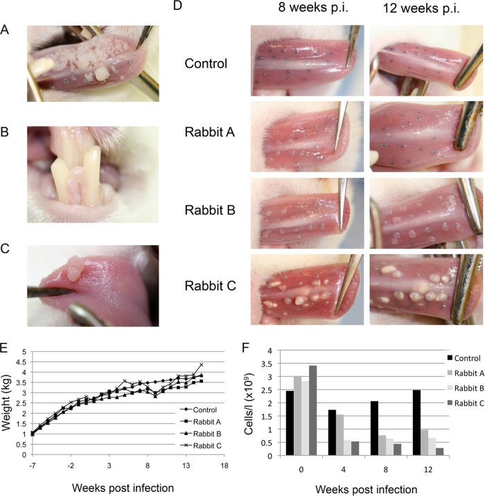
FIG 1.
(A) Presence of florid ROPV-induced warts in immunosuppressed rabbits at 15 weeks postinfection. In immunocompetent rabbits, lesions were not detectable beyond 10 weeks. (B and C) In the immunosuppressed rabbits, secondary infections were also apparent at nonscarified sites by 15 weeks postinfection (p.i.). (D) Lesion size and abundance varied among immunosuppressed rabbits (rabbits A, B, and C), depending on the extent of immunosuppression (see panel F). Immunocompetent rabbits developed much smaller lesions (control), with tattoo marks indicating the sites of previous infection by week 12. (E) The cyclosporine-dexamethasone immunosuppression regimen was found not to have a major effect on weight gain in juvenile (growing) rabbits during the time course of the immunosuppression experiment. (F) To establish the extent of immunosuppression, T cell counts of the three immunosuppressed rabbits shown in panel D are shown as gray columns (rabbits A, B, and C); T cell counts of a typical immunocompetent animal are shown as black columns.

