FIG 2.
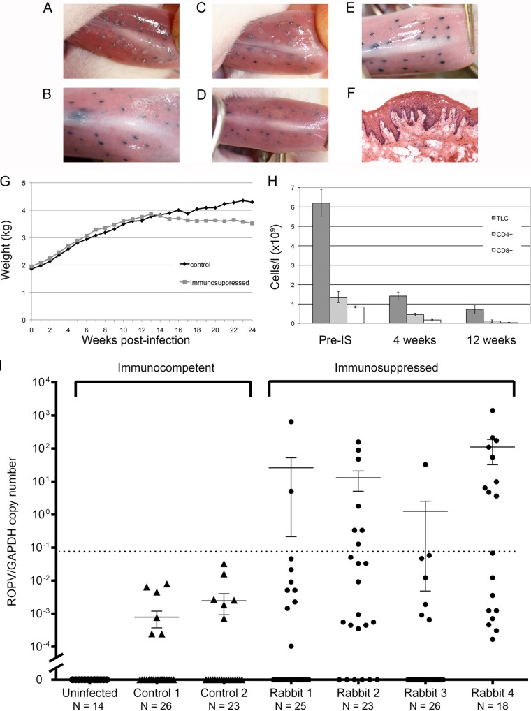
(A to D) Typical appearance of tattoo-marked tongue sites in two ROPV-infected rabbits at 4 weeks postinfection (A and C) and 12 weeks postinfection (B and D). At 4 weeks postinfection (A and C), papillomas are apparent overlying tattoo-marked infection sites. At 12 weeks postinfection in the absence of immunosuppression (B and D), only tattoo marks remain at previous sites of infection. (E) The appearance of tattoo marks in the uninfected control rabbit in which no papilloma lesions developed during the course of the study are indistinguishable from the regressed sites shown in panels B and D. (F) Histology of a single microlesion (with typical papilloma features) detected in rabbit 2 at 12 weeks postimmunosuppression. (G) In adult rabbits, the cyclosporine-dexamethasone immunosuppression regimen generally caused weight loss, with rabbits being culled when their weight loss exceeded 20% of their total body weight. The graph shown is typical of what was seen in repeated experiments. (H) Decline in total lymphocyte counts (TLC; dark gray) and CD4- and CD8-positive (light gray/white) lymphocyte levels following the onset of immunosuppression (IS) in the adult infected rabbits shown in panel I. (I) Numbers of ROPV copies per copy of GAPDH DNA at sites of previous infection in postregression control rabbits (triangles) and postregression immunosuppressed rabbits (circles). Cyclosporine-dexamethasone was administered at week 12 to the immunosuppressed animals, and viral copy numbers were measured at individual tattoo-marked sites at week 24. ROPV/GAPDH copy numbers below the dotted line are those that are typically observed in rabbits with a latent infection or where no ROPV is detectable (12). Elevated copy numbers were seen only at tattoo-marked sites in the immunosuppressed animals.

