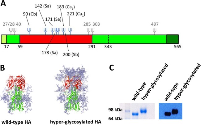
FIG 1.
Covering antigenic sites of HA with N-linked glycosylation. (A) Schematic of the influenza HA and the position of introduced glycans. The signal peptide is shown in light green, the head and stalk domain of the ectodomain in red and green, respectively, and the transmembrane and cytoplasmic domains are depicted in dark green. Glycans indicated in gray are present in the wild-type HA. Additional glycans in the hyperglycosylated HA are depicted in blue. (B) Modeling of introduced glycans on the crystal structure of the PR/8 HA (PDB 1RU7). Original and additional glycans were modeled on the crystal structure of the PR/8 HA using GlyProt (37) and drawn using PyMol (DeLano Scientific). Because of modeling limitations, only 6 out of the 7 added potential N-linked glycans are present on the hyperglycosylated HA model. (C) The ectodomain of the wild-type HA and the hyperglycosylated HA containing the T4-foldon trimerization domain and a His tag were expressed in 293T cells, and purified glycoproteins were run on an SDS-PAGE polyacrylamide gel under denaturing and reducing conditions. A clear increase in molecular weight is present for the hyperglycosylated HA compared to the wild-type HA, indicating the presence of additional glycosylation.

