
FIG 3.
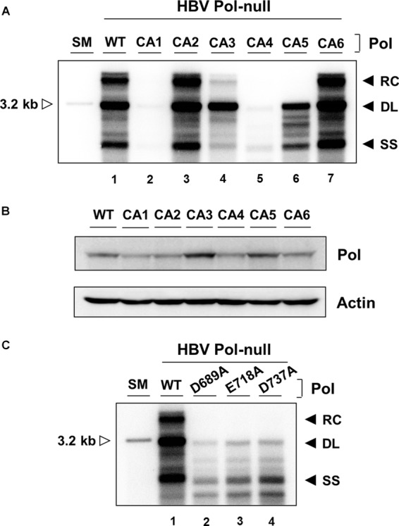
Analysis of CA mutants for the ability to support viral genome replication. (A) Cells were cotransfected with a HBV P-null replicon construct along with WT Pol or individual CA mutant constructs as indicated. Intracellular capsid-associated DNAs were isolated and analyzed by Southern blotting. The viral replication intermediates, RC, DL, and SS DNAs, are denoted. A restriction fragment representing one HBV genomic unit (3.2 kb) serves as a size marker (SM) (open arrowhead). The experiment was repeated four times, and representative data are shown. (B) Cytoplasmic lysates, equivalent to 3% input from the same cells transfected with the constructs indicated, were analyzed by immunoblotting. The levels of HBV Pol and β-actin were measured using anti-Flag and anti-actin antibodies, respectively. (C) Intracellular capsid-associated DNAs obtained from three catalytic-site mutants were isolated and analyzed similarly to the assay described above.