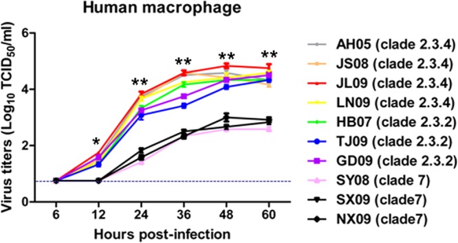
FIG 4.

Viable virus output from macrophages infected with clade 2.3.4 (AH05, JS08, JL09, and LN09), clade 2.3.2 (HB07, TJ09, and GD09), or clade 7 (SY08, SX09, and NX09) virus at an MOI of 0.001. At 6, 12, 24, 36, 48, and 60 h postinfection, virus titers in the supernatants were determined by TCID50 assays on MDCK cells. The values are expressed as means ± standard deviations (SD) (n = 3). *, P < 0.05, and **, P < 0.01, between clade 2.3.4-virus infected cells and clade 7 virus-infected cells. The dashed line represents the detection limit of the TCID50 assay.
