FIG 5.

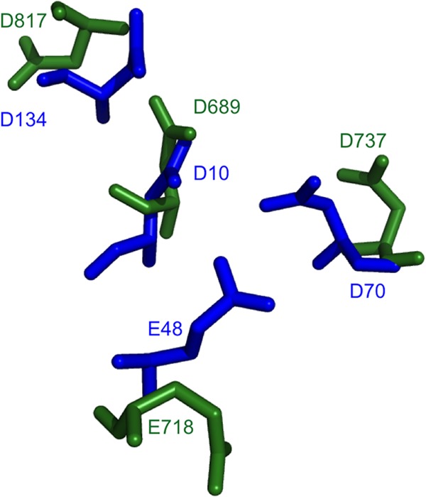
Superimposition of residues D689, D737, D817, and E718 in our model with the corresponding residues in the E. coli structure. HBV and E. coli RNase H residues are shown by blue and green sticks, respectively.

Superimposition of residues D689, D737, D817, and E718 in our model with the corresponding residues in the E. coli structure. HBV and E. coli RNase H residues are shown by blue and green sticks, respectively.